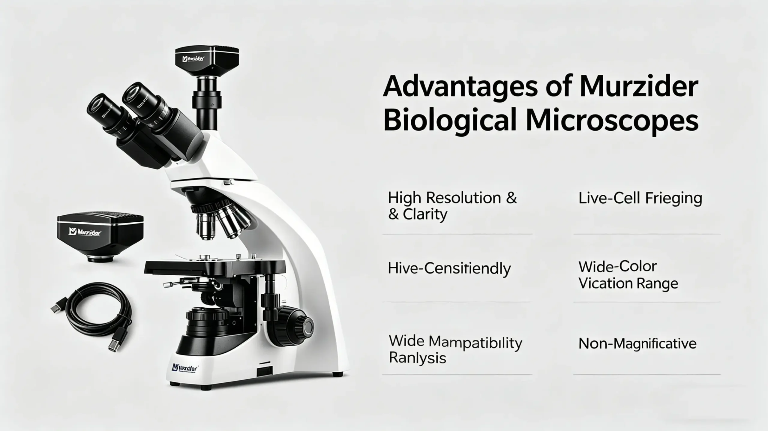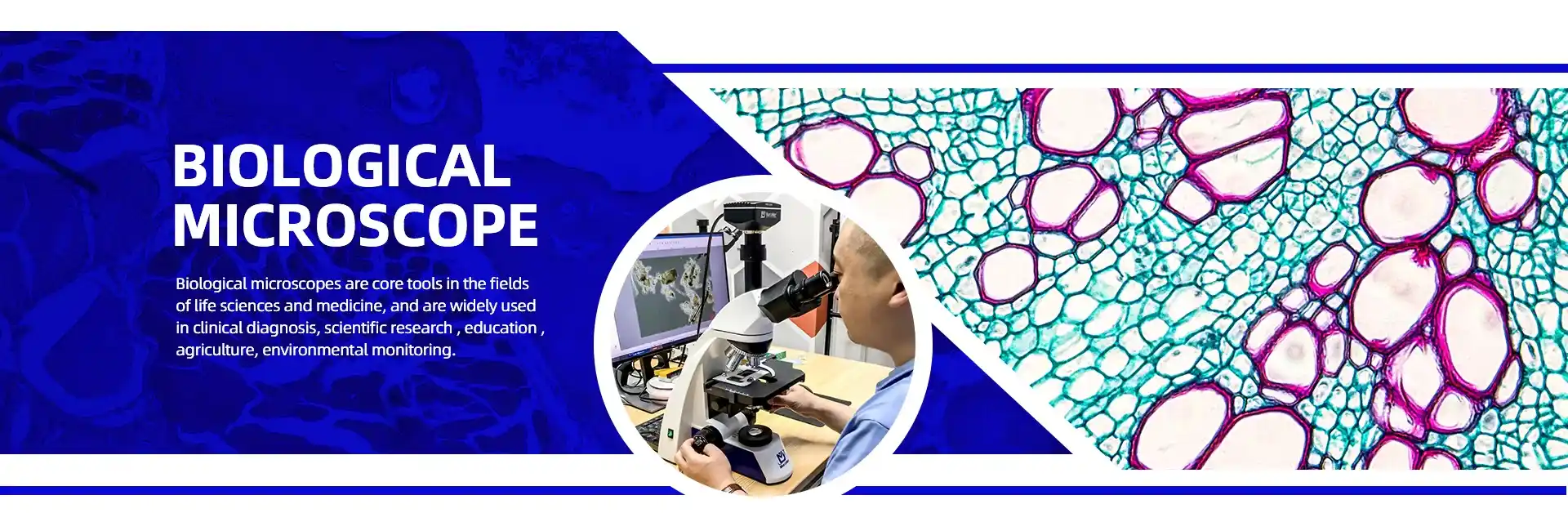
Biological Microscopes for Education & Clinical Labs
Murzider Biological Microscopes deliver reliable, high-performance optical solutions built for daily use in education and clinical laboratories.
Engineered for clarity, durability and consistent performance, our microscopes empower teaching excellence, accurate clinical diagnostics, and precise laboratory observations every single day.
- Deliver clear, crisp imaging for classroom teaching & clinical testing
- Stable 40X–1000X magnification for routine lab observations
- Versatile brightfield & phase contrast for education & clinical samples
- High-quality optical system ensures accurate sample identification
- Ergonomic, low-maintenance design for long-term daily use
Working Principle
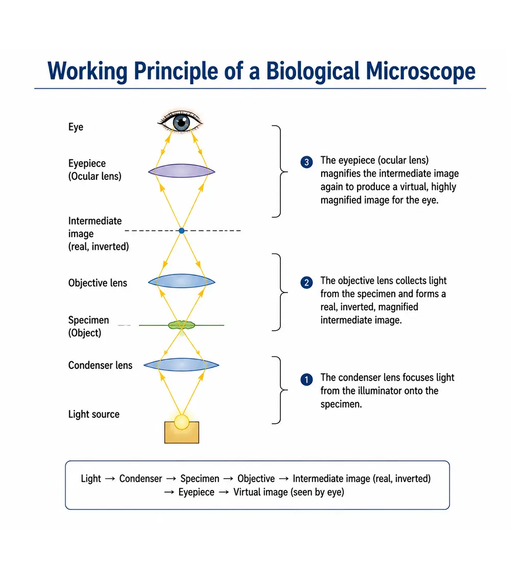
Murzider biological microscopes use transmitted-light optics:
1. Light source emits illumination from below the sample
2. Condenser lens focuses light evenly through the specimen
3. Objective lens magnifies the sample and forms primary image
4. Light carries image through optical path to eyepiece/camera
5. Eyepiece further magnifies image for observation
6. Total magnification = Objective × Eyepiece (typically 40X–1000X)
7. Clear details of cells, tissues, and microorganisms revealed
Key Components
Objective Lenses
4x, 10x, 40x, 100x (oil immersion) high-NA plan-achromatic lenses for exceptional clarity and resolution.
Illumination System
LED/halogen transmitted-light source with adjustable brightness for consistent, cool illumination.
Condenser & Diaphragm
Abbé condenser with iris diaphragm controls light intensity and contrast for optimal sample visualization.
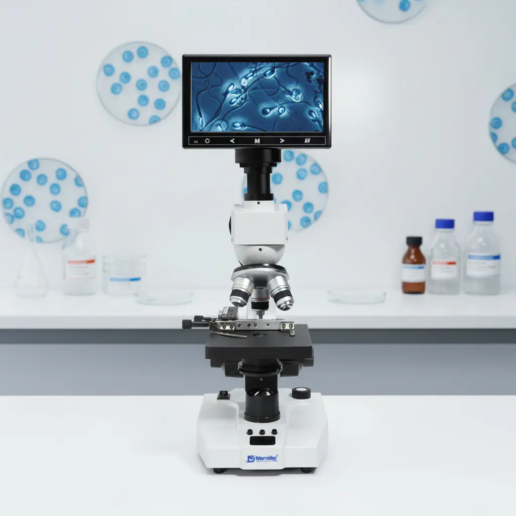
Eyepiece (Ocular)
Wide-field 10x eyepieces (binocular/trinocular) for comfortable viewing; trinocular supports digital cameras.
Mechanical Stage
Precision XY-controlled stage with slide holder for accurate sample positioning and scanning.
Focus System
Coarse & fine focus knobs for smooth, precise focusing to reveal fine cellular details.
Main Applications
Cell Biology
Observe cell structure, division, morphology, and live-cell dynamics in cultures and tissue samples.
Histology & Pathology
Analyze stained tissue sections for disease diagnosis, cancer screening, and histological research.
Microbiology
Identify bacteria, fungi, parasites, and microbes; study morphology and growth patterns.
Education & Training
Teaching tools in schools/universities for biology, anatomy, and life science laboratory training.
Clinical Diagnostics
Blood analysis, urine testing, and pathogen identification in medical laboratories.
Biotechnology & Research
Drug development, genetic research, stem cell studies, and biotech sample analysis.