Murzider Fluorescence Microscopes
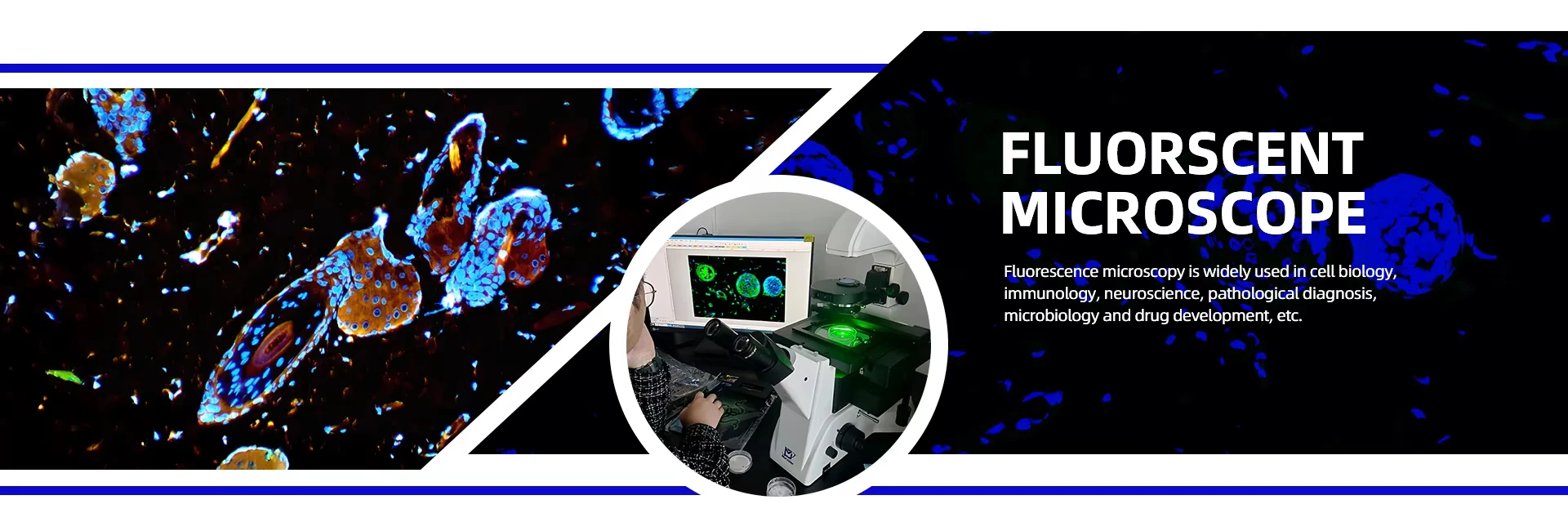
Murzider fluorescence microscopes are engineered for high-precision laboratory imaging, ideal for cell biology, medical research, neuroscience and material science applications.
Our fluorescence microscope series adopts optimized optical structure and high-quality filter sets, delivering high contrast, clear resolution and stable performance for long-term professional use.
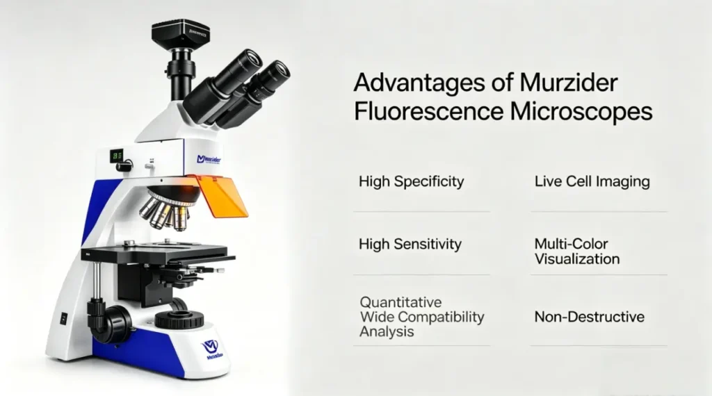
- Highly specific target molecule imaging
- Ultra-high sensitivity for tiny structure observation
- Non-invasive detection for living biological samples
- Supports multi-channel fluorescence imaging
Working Principle of Fluorescence Microscopes
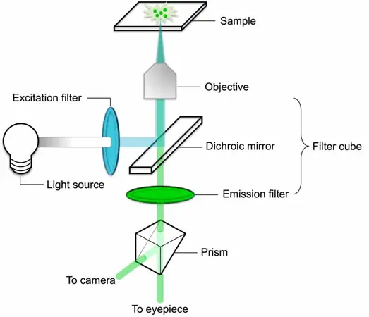
Murzider fluorescence microscopes operate on the principle of fluorescence excitation and emission:
1. A light source emits high-intensity excitation light
2. The excitation filter selects only the specific wavelength needed to excite the fluorophore
3. The dichroic mirror reflects the excitation light to the sample and transmits emitted light
4. Fluorophores in the sample absorb light and emit longer-wavelength fluorescence
5. Emission filter blocks unwanted excitation light and passes only fluorescence to the detector
6. The objective lens captures the fluorescent signal to form a clear image
Key Components of Murzider Fluorescence Microscopes
Light Source
High-power mercury lamps, xenon lamps, or LEDs provide the intense excitation light required to stimulate fluorescence in sample molecules.
Filter Sets
Precision optical filters select specific excitation wavelengths and block unwanted light while allowing emitted fluorescence to pass through.
Objective Lens
High-resolution lenses collect fluorescence from the sample and focus it to create detailed, magnified images with exceptional clarity.
Dichroic Mirror
Special optical mirror that reflects excitation light toward the sample and transmits fluorescence to the detection system.
Detector System
High-sensitivity cameras or eyepieces capture and display fluorescent images for observation, analysis, and documentation.
Stage & Sample Holder
Stable platform for positioning samples, often with precision controls for accurate focusing and scanning.
Applications of Murzider Fluorescence Microscopes
Cell Biology
Visualize cell structures, organelles, protein localization, and dynamic cellular processes in living and fixed cells.
Medical Research
Study disease mechanisms, identify pathogens, analyze tissue samples, and develop new diagnostic techniques.
Neuroscience
Image neural networks, track brain activity, study synaptic connections, and observe neurological disorders.
Genetics
Map gene expression, locate specific DNA sequences, analyze chromosome structure, and study genetic disorders.
Material Science
Characterize nanomaterials, analyze polymer structures, and study advanced material properties at micro levels.
Pharmaceutical Development
Screen drug candidates, analyze drug delivery systems, and evaluate treatment effects at cellular levels.